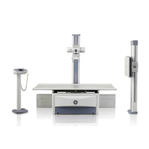
Set up exams quickly and confidently. Position patients and devices fast and flexibly. Edit images and transfer exams easily and effortlessly. Keep patients comfortable and cared for. At every step of every exam, the Optima XR646 is designed to simplify, shorten, and bring more satisfaction to every technologist’s workday.
This system lets you position yourself for an easier experience, extend your performance clinically and financially, and see the excellence in every study.
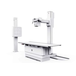
The Optima XR646’s striking image quality is designed to support accurate diagnoses, confident decisions, patient satisfaction, and frequent referrals.
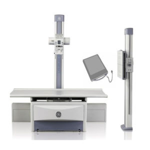
- Diversify your detector: Share the GE FlashPad wireless detector among all of your compatible GE X-ray products (including mobile systems) for added productivity.
- Turn data into dose efficiency: The Detector Exposure Index represents the ratio of the received exposure at the detector surface and the predefined expected exposure. DEI gives you the ability to monitor variance in image quality and dose and determine “dose creep” for fixed techniques. It also provides the ability to determine average dose by anatomy, view and patient size. Dose data can also be used to establish consistency in protocols and standardize image quality room to room and site to site.
- Dual-Energy Subtraction: See bone and soft tissue better: Separate soft tissue and bone structures by making one acquisition with two exposures at different energy levels in less than 200 milliseconds. Three images are generated providing three separate views of the chest: a standard radiographic image, an image with the bones “subtracted,” and an image of just the bones to highlight foreign objects or calcified structures.
- Auto Image Paste : Get seamless long-bone or full spine images in one fast, low-dose exam: Automatically acquire multiple overlapping images of larger anatomy such as the entire spine at the wall stand.